Female Internal Anatomy / Anatomy Of The Female Reproductive System / Even though the uterus is primarily a pelvic organ, but during later stages of pregnancy due to hypertrophy and.
Female Internal Anatomy / Anatomy Of The Female Reproductive System / Even though the uterus is primarily a pelvic organ, but during later stages of pregnancy due to hypertrophy and.. It is a highly muscular, childbearing organ in females, approximating 3 x 2 x 1 inches in size in a nulliparous. At the level of the pelvic bones, the abdomen. The female reproductive anatomy includes both external and internal structures. In virgins, the hymen usually encircles the opening like a tight ring, but it may completely cover the opening. The uterus or womb accommodates the embryo which develops into the foetus.
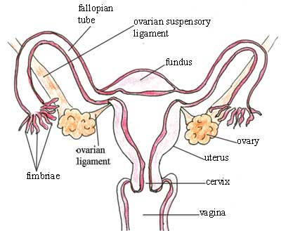
The detailed structure depends on a lot of factors such as the dog breed, age, and weight. The uterus or womb accommodates the embryo which develops into the foetus. Media in category female human anatomy the following 144 files are in this category, out of 144 total. An anatomically female's internal reproductive organs are the vagina, uterus, fallopian tubes, cervix, and ovary. The area containing these organs is called the vulva.
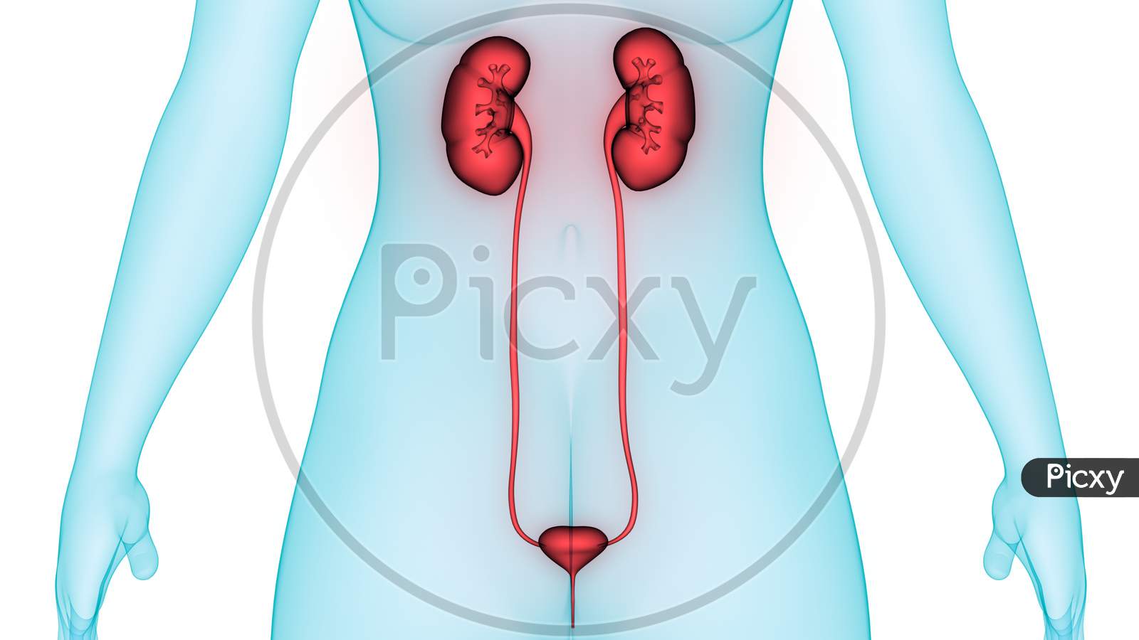
Image Of Female Internal Organs Kidneys With Bladder Anatomy Io729522 Picxy from images.picxy.com The function of the external female reproductive structures (the genitals) is. Even though the uterus is primarily a pelvic organ, but during later stages of pregnancy due to hypertrophy and. At the level of the pelvic bones, the abdomen. An anatomically female's internal reproductive organs are the vagina, uterus, fallopian tubes, cervix, and ovary. Anatomy is a branch of biology and medicine that studies the morphology and structure. Female internal organs, by vesalius. The area containing these organs is called the vulva. The uterus or womb accommodates the embryo which develops into the foetus.
The function of the external female reproductive structures (the genital) is twofold:
What parts make up the female anatomy? The mons pubis is a rounded mound of fatty tissue that covers the pubic bone. The female reproductive system is an intricate arrangement of structures that can separate into external and internal genitalia. The internal anatomy is all of the parts you can't see, and where the reproductive magic happens. Low key lighting during a spay surgery at a vet clinic in london ontario canada. This diagram depicts anatomy female 1024×1111 with parts and labels. At the level of the pelvic bones, the abdomen. Female anatomy of internal organs with skeleton, rear and front views. The abdomen (commonly called the belly) is the body space between the thorax (chest) and pelvis. The area containing these organs is called the vulva. It is a highly muscular, childbearing organ in females, approximating 3 x 2 x 1 inches in size in a nulliparous. The function of the external female reproductive structures (the genital) is twofold: The uterus or womb accommodates the embryo which develops into the foetus.
This diagram depicts anatomy female 1024×1111 with parts and labels. Fallopian tubes and ovaries form the adnexa of the uterus. The uterus (womb) is the site of implantation of a fertilized ovum, growth and development of the fetus during pregnancy and labor.during reproductive cycles when implantation does not occur, the uterus is the source of menstrual flow. Get prepared for your anatomy exams: The mons pubis is a rounded mound of fatty tissue that covers the pubic bone.
Female Human Anatomy Internal Organs Diagram Physiology Structure Medical Profession Morphology Healthy Stock Images Page Everypixel from st4.depositphotos.com Abdominal and thoracic female organ set, realistic heart, lungs, stomach, liver, kidneys, spleen, large and back side view of male skeleton with kidneys. Illustrated sagittal view of the female reproductive system. Media in category female human anatomy the following 144 files are in this category, out of 144 total. 3d art illustration of anatomy back side view of male skeleton with kidneys Even though the uterus is primarily a pelvic organ, but during later stages of pregnancy due to hypertrophy and. Browse 38,615 female anatomy stock photos and images available, or search for female anatomy illustration or male female anatomy to find more great stock photos and pictures. It is a highly muscular, childbearing organ in females, approximating 3 x 2 x 1 inches in size in a nulliparous. The internal anatomy is all of the parts you can't see, and where the reproductive magic happens.
Anatomy is a branch of biology and medicine that studies the morphology and structure.
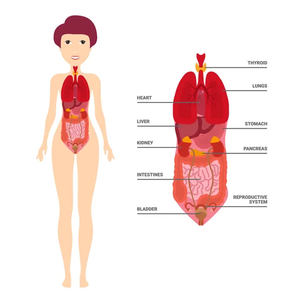
Human body internal organs, skeleton, skeletal bones, circulatory cardiovascular system. Female figure with select internal anatomy. Anatomy is a branch of biology and medicine that studies the morphology and structure. Media in category female human anatomy the following 144 files are in this category, out of 144 total. Shown are the clavicle, heart and kidneys. Anatomy of internal organs female : Fallopian tubes and ovaries form the adnexa of the uterus. Female internal organs, by vesalius. It is a highly muscular, childbearing organ in females, approximating 3 x 2 x 1 inches in size in a nulliparous. At the level of the pelvic bones, the abdomen. The diaphragm forms the upper surface of the abdomen. Whether or not a baby is present, the hormonal functions that come with your internal anatomy impact your everyday life. An anatomically female's internal reproductive organs are the vagina, uterus, fallopian tubes, cervix, and ovary.
It is a highly muscular, childbearing organ in females, approximating 3 x 2 x 1 inches in size in a nulliparous. Anatomy is a branch of biology and medicine that studies the morphology and structure. Female internal anatomy back view stock illustration illustration of organs female 20085552 : Media in category female human anatomy the following 144 files are in this category, out of 144 total. An illustration showing a theory of vision published in treatise on man by rene descartes.
Olcreate Heat Anc Et 1 0 Antenatal Care Module 3 Anatomy And Physiology Of The Female Reproductive System 3 4 1 Fallopian Tubes And Ovaries from www.open.edu Anatomy is a branch of biology and medicine that studies the morphology and structure. The internal anatomy is all of the parts you can't see, and where the reproductive magic happens. Female figure with select internal anatomy. Thank you for visit anatomynote.com. In humans, the female reproductive system is immature at birth and develops to maturity at puberty to be able to produce gametes, and to carry a foetus to full term. The mons pubis is a rounded mound of fatty tissue that covers the pubic bone. The function of the external female reproductive structures (the genital) is twofold: 3d art illustration of anatomy back side view of male skeleton with kidneys
At the level of the pelvic bones, the abdomen.
3d art illustration of anatomy back side view of male skeleton with kidneys The female reproductive system is an intricate arrangement of structures that can separate into external and internal genitalia. The area containing these organs is called the vulva. Young female teacher in biology class, holding digital tablet and teaching human body anatomy, using artificial body model to explain internal organs. The uterus (womb) is the site of implantation of a fertilized ovum, growth and development of the fetus during pregnancy and labor.during reproductive cycles when implantation does not occur, the uterus is the source of menstrual flow. The internal anatomy is all of the parts you can't see, and where the reproductive magic happens. Female internal organs, by vesalius. Abdominal and thoracic female organ set, realistic heart, lungs, stomach, liver, kidneys, spleen, large and back side view of male skeleton with kidneys. An illustration showing a theory of vision published in treatise on man by rene descartes. Fallopian tubes and ovaries form the adnexa of the uterus. Female anatomy of internal organs with skeleton, rear and front views. Female figure with select internal anatomy. Get prepared for your anatomy exams:
Even though the uterus is primarily a pelvic organ, but during later stages of pregnancy due to hypertrophy and female internal. Human body internal organs, skeleton, skeletal bones, circulatory cardiovascular system.


0 Komentar